1601006029 LONG CASE
Case presentation(1601006029)
CASE HISTORY
A 65 year old female from Alingapuram Came to opd 25 days back with complaints of fever & swelling in bilateral lower limbs since 1 week and Shortnessof breath since 4 days
History of present illness:
The patient was apparently asymptomatic 25 days ago then she developed:
SOB ( shortness of breath )- while sitting and was insidious in onset, gradually progressive ( Grade IV )
Swelling in the legs- started bilaterally in the feet and gradually progressed upto mid calf level ( Grade 2 ) associated with cough.
Associated with fever which is of low grade and continuous type
No h/o chest pain , palpitations, decreased urine output , blood in urine, burning micturition.
Past History:
No history of similar complaints in the past.
Patient is a known case of Hypertension since 2 years for which she took medication irregularly.
Not a known case of Diabetes Mellitus, Asthma, TB, Epilepsy
H/o b/l knee pain since 1 year for which she was prescribed pain killers ( mostly NSAIDS )
No significant surgical history.
Family History: Not significant
Personal History:
Diet: Mixed
Appetite : Decreased
Sleep: Reduced
Bowel and Bladder: regular
Addictions: none
Drug History: No known drug allergies.
General Examination:
The conscious, coherent, not co operative.
Moderately built, moderately nourished.
Pallor- present
Edema- pitting type edema
(grade II ) involving both feet and legs upto mid calf.


Icterus- absent
Cyanosis : absent
Clubbing : absent
Koilonychia: absent
Lymphadenopathy: absent
Vitals:
Temperature: afebrile
Pulse Rate: 82 beats/min
Blood pressure: 130/ 80 mm hg
Respiratory Rate: 20 cycles/min
Spo2: 98%
Systemic examination:
Respiratory System:
Shape of chest - normal
Inspection of upper respiratory system-
• oral cavity- normal
• Nose- normal
• Pharynx- normal
Lower Respiratory Tract:
Inspection:
• trachea: central
• Symmetry of chest : symmetrical
• Movement: B/L symmetrical expansion of chest respiration


• No scars, engorged veins or sinuses.
Palpation:
All inspectory findings are confirmed by palpation.
• Trachea: central - confirmed by three finger test.

• Assessment of anterior and posterior chest expansion- B/L symmetrical expansion of chest.
• No chest wall tenderness
• Vocal fremitus- decreased in inframammary , infra axillary and infra scapular
Percussion : done in sitting position
Right Left
Supraclavicular Resonant Resonant
Infraclacicular Resonant Resonant
Mammary Resonant Resonant
Inframammary. stony dull stony dull
Axillary. Resonant Resonant
Infraaxillary stony dull stony dull
Supra scapular Resonant Resonant
Interscapular Resonant Resonant
Infrascapular stony dull stony dull
Auscultation:
Right Left
Supraclavicular NVBS NVBS
Infraclacicular NVBS NVBS
Mammary NVBS NVBS
Inframammary diminished diminished
Axillary NVBS NVBS
Infraaxillary diminished. Diminished
Supra scapular NVBS NVBS
Interscapular NVBS NVBS
Infrascapular diminished diminished
Vocal resonance - markedly diminished over the fluid area
Basal crackles are present.

Cardiovascular System :
Inspection :
- No scars sinuses and engorged veins
- No visible pulsations
Palpation:
- apical impulse : palpable in fifth inter coastal space
Auscultation:
• S1 and S2 heard
• No murmurs
Abdomen:
Inspection:
- shape elliptical
- Quadrants of abdomen moving in accordance with respiration.
- Umbilicus- central and inverted
- No scars sinuses or engorged veins
Palpation:
• No tenderness
• No organomegaly
Percussion
• tympanic
Auscultation:
• Normal bowel sounds heard
CNS:
• Higher mental functions-normal
• Cranial nerves- intact
• Sensory system- normal
• Motor system- normal
• Cerebellar signs- absent
Investigations:
1.Complete blood picture
• Hb 6.2 g/dl

2.Complete Urine examination
• albumin +2
3.Liver function Tests

4.RBS -82
5.Renal function Tests

• Raised urea- 94 mg/dl
• Raised creatinine- 6.3 mg/dl
• Raised uric acid - 9.9 mg/dl
X-Ray

Ultrasound
- few subcentrimetric anechoic cyst in b/l kidneys
- Rt kidney- CMD lost. Grade III RPD
- Lt kidney- CMD partially maintained. Grade II RPD

ECG

Rate - 100b/min
Normal sinus rhythm
Normal axis
P-wave ,qrs complex , t wave , pr interval ,st segment all appears to be normal.
Provisional Diagnosis: Renal failure with bilateral pleural effusion
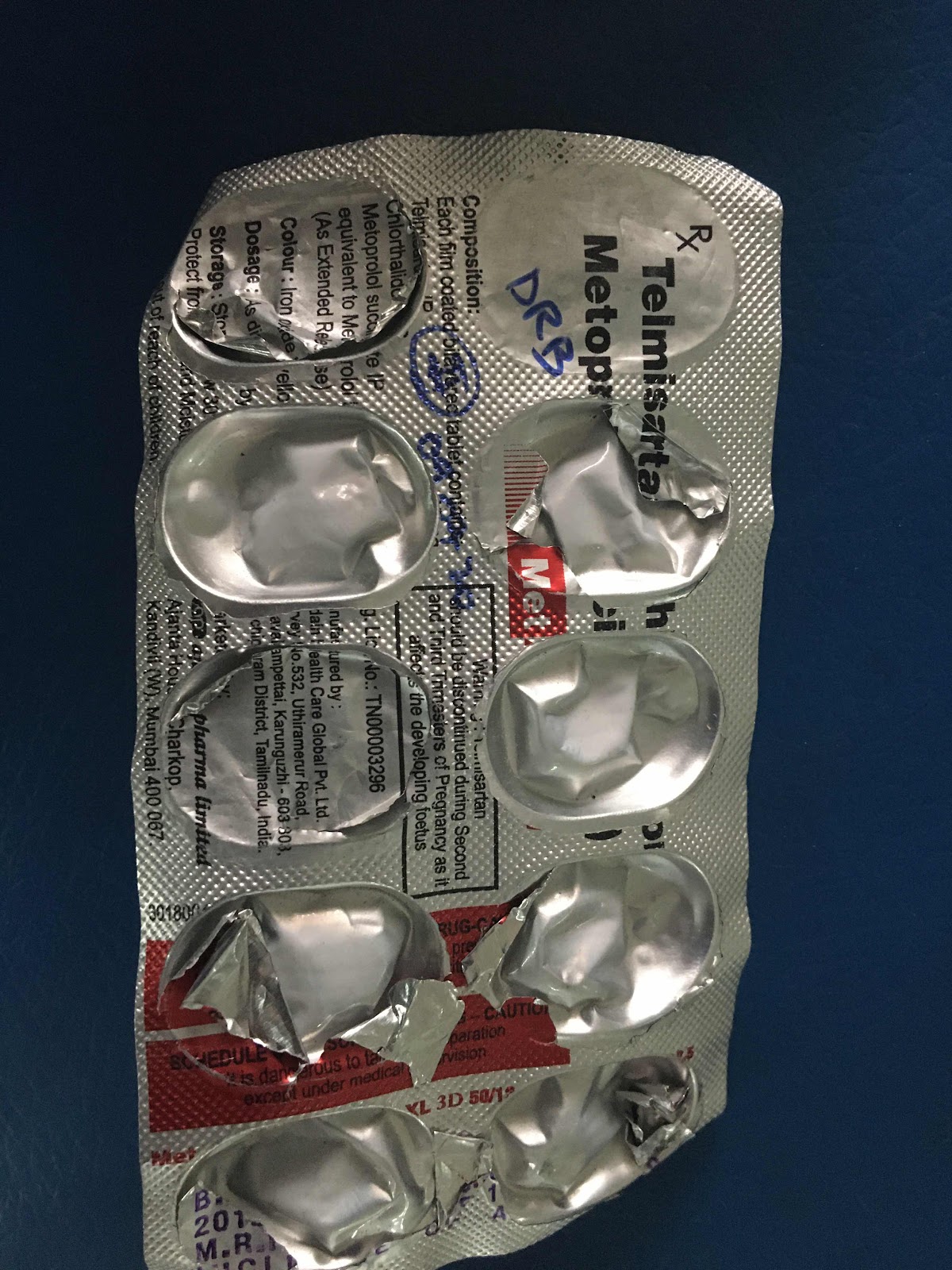
Treatment History:
• Inj piptaz 2.25 g iv TID
• TAB Lasix 40 mg PO/ BD
• TAB Nodosis 500 mg PO/BD
• TAB Orofer XT PO/OD
• TAB Shelcal CT PO/OD
• Inj Erythropoietin 4000 IU SC- twice weekly
• Salt and fluid Charting
• Vitals monitor and strict input/output charting.


Dialysis:
Patient underwent 6 sessions of dialysis since admission


Comments
Post a Comment